Comprehensive skin checks
Systematic examination guided by your history, risk factors and areas of concern.
Accredited skin cancer care · Tamworth, NSW
Comprehensive skin checks, dermoscopic assessment, diagnosis and surgical management—supporting patients across Tamworth, rural and regional New South Wales.
Appointments are provided through MY GP Hub – West Tamworth.
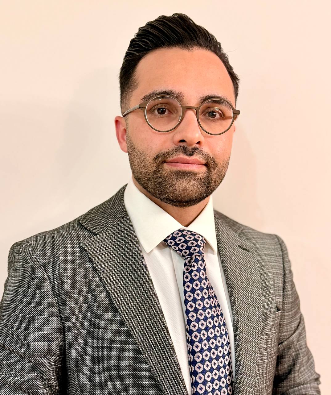
Dr Amin Amiri is an Accredited Skin Cancer Doctor with the Skin Cancer College Australasia. This means that he has successfully completed a high level of training with the College and has proven skills in the diagnosis, treatment and management of skin cancer.
Local care. Considered decisions.
Dr Amin Amiri is a General Practitioner and an Accredited Skin Cancer Doctor with the Skin Cancer College Australasia. He holds a Master of Medicine in Skin Cancer from the University of Queensland.
His work encompasses early detection, dermoscopic evaluation, biopsy, definitive treatment and follow-up. The aim is to bring accessible, evidence-based skin cancer care closer to people living in Tamworth and surrounding rural communities.
Each plan is based on the lesion, pathology, anatomical site, patient health and personal preferences.
Systematic examination guided by your history, risk factors and areas of concern.
Detailed dermoscopic assessment, clinical photography and surveillance where appropriate.
Punch, shave or excisional biopsy selected according to the lesion and clinical circumstances.
Treatment of suitable BCC, SCC and melanoma, planned for function, healing and appearance.
Reconstructive options for selected surgical defects, including local flaps and full-thickness grafts.
Close collaboration with dermatology and plastic surgery for complex or difficult cases.
Dermoscopy
Dermoscopy is a simple, painless examination that uses magnification and specialised light to reveal colours and structures within a skin lesion that cannot be seen clearly with the naked eye.
Used together with clinical history and examination, it improves assessment of both pigmented and non-pigmented lesions and helps determine whether a spot should be monitored, biopsied or treated.
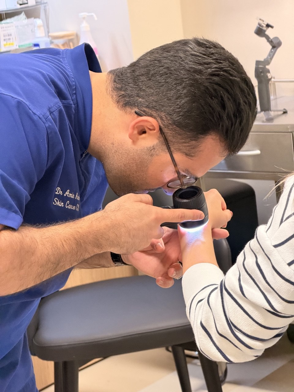
Subtle patterns may help identify suspicious change before it becomes obvious to the naked eye.
Dermoscopy helps select lesions that need biopsy while supporting safe monitoring of appropriate lesions.
Images can be recorded and compared over time to assess meaningful change during surveillance.
Dermoscopy supports clinical decision-making but does not replace histopathology. When a lesion remains suspicious, biopsy is required for a definitive diagnosis.
Common skin cancers
Often slow-growing, but early assessment can simplify treatment and reconstruction.
May require prompt diagnosis and treatment, particularly at high-risk sites.
Early recognition and appropriate excision are central to achieving the best possible outcome.
Total body mapping
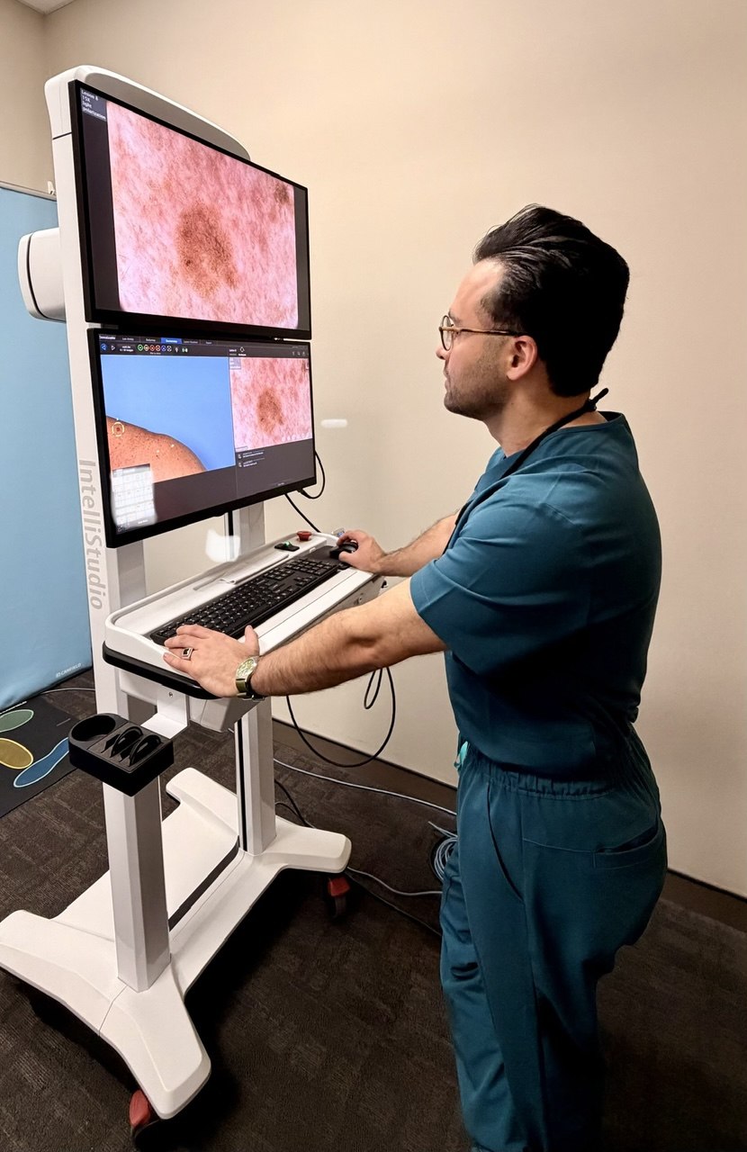
Total body photography creates a detailed visual record of the skin. Combined with digital dermoscopy, it helps identify new lesions and subtle changes in existing moles during follow-up.
Standardised photographs provide a reference for future comparison.
Selected lesions can be recorded at high magnification for targeted monitoring.
Particularly useful for selected patients with many or atypical moles, or a personal or family history of melanoma.
Total body mapping supports—but does not replace—a clinical skin examination. Suitability and follow-up intervals are determined individually.
Book total body mappingPatient education
This overview explains the usual clinical pathway. The appropriate investigation and treatment depend on the individual lesion, its location, pathology and your general health.
Diagnosis
The skin is examined in the context of your personal and family history. Dermoscopy allows structures beneath the surface to be assessed more closely.
If a lesion is suspicious, tissue is removed under local anaesthetic. Complete excision biopsy is generally preferred for suspected melanoma when practical.
A pathologist examines the tissue under a microscope. The report confirms the diagnosis and records features that guide staging and further treatment.
Sampling
A circular instrument removes a small, full-thickness core of skin. It is useful for sampling selected lesions or inflammatory skin conditions and may require a stitch.
A blade removes a superficial or saucer-shaped sample. The depth is chosen for the suspected diagnosis and the specimen is sent to pathology.
The entire visible lesion is removed with a narrow margin and closed with stitches when possible. This is generally preferred when melanoma is suspected.
The biopsy method is selected by the clinician. A partial biopsy may be appropriate for a very large lesion, a sensitive anatomical site, or where complete removal would affect function.
Types
The most common skin cancer. It usually grows locally and slowly but can damage surrounding tissue if untreated.
A keratinocyte cancer that can grow more quickly. Some SCCs have a meaningful risk of spreading.
A cancer of melanocytes. Early diagnosis is important because melanoma can spread to lymph nodes and other organs.
A rare, aggressive skin cancer that generally requires prompt specialist and multidisciplinary management.
Management
Treatment is selected after considering the pathology, tumour size and depth, anatomical site, risk of recurrence, patient health and treatment preferences.
Standard excision is commonly used. Selected cancers may require margin-controlled surgery, wider excision, a local flap or a skin graft.
For carefully selected superficial or low-risk lesions, options may include topical medicines, curettage and cautery, cryotherapy, photodynamic therapy or radiotherapy.
Management may involve dermatology, plastic surgery, surgical oncology, radiation oncology or medical oncology. Immunotherapy or targeted treatment may be appropriate for some advanced cancers.
Mohs surgery removes a skin cancer one thin layer at a time. Each layer is examined under a microscope while the patient waits, and further tissue is removed only where cancer cells remain. This continues until the examined margins are clear.
Because the technique checks the surgical margin during the procedure, it can preserve as much healthy tissue as possible. It may be considered for selected high-risk, recurrent or poorly defined skin cancers, particularly in functionally or cosmetically important areas such as the face.
Mohs surgery is performed by appropriately trained specialist surgeons. Dr Amin Amiri works closely with Mohs surgeons and can arrange referral and coordinate ongoing care when this approach is considered appropriate.
Sun damage & prevention
Actinic keratoses—also called solar keratoses or sunspots—often feel rough or scaly and commonly occur on frequently exposed areas such as the face, scalp, ears, forearms and backs of the hands. They develop after repeated ultraviolet exposure and indicate that the surrounding skin has also sustained sun damage.
Most do not become cancer, but some can progress to squamous cell carcinoma. A lesion that becomes thicker, tender, rapidly enlarging, bleeding or persistent should be reassessed and may require biopsy.
Treatment options
Liquid nitrogen freezes and destroys abnormal cells. It is often used for individual, clearly defined actinic keratoses. Temporary stinging, redness, blistering, crusting or a pale mark can occur while the area heals.
A prescription chemotherapy cream used to treat an area of sun-damaged skin rather than only one visible spot. Redness, inflammation, crusting and discomfort are expected during treatment and should be monitored by your clinician.
A prescription immune-response cream used for selected actinic keratoses and some superficial skin cancers. It stimulates a local immune reaction and commonly causes redness, irritation and crusting in the treated area.
Topical creams must be used only on the area and schedule prescribed by your doctor. Suspicious lesions may need biopsy before field treatment begins.
Make sunscreen a habit
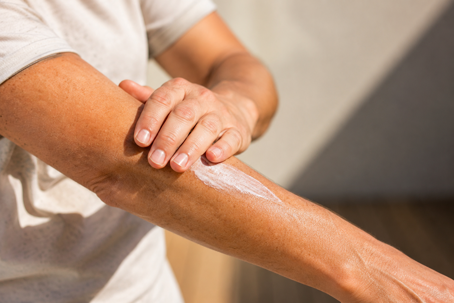
Use SPF50+ broad-spectrum, water-resistant sunscreen on exposed skin. Apply it generously 20 minutes before going outdoors and reapply at least every two hours—more often after swimming, sweating or towelling.
Sunscreen does not block all UV radiation. Combine it with protective clothing, a broad-brimmed hat, sunglasses and shade whenever the UV Index is 3 or above.
Read SunSmart sunscreen guidanceTrusted resources
Melanoma information, research, treatment and patient resources.
Australasia’s peak body for primary-care skin cancer practitioners.
Australian general-practice guidance on skin cancer risk and skin checks.
National cancer information, prevention guidance and support services.
Specific information about skin cancer symptoms, diagnosis and treatment.
History, full skin examination when indicated, and dermoscopic evaluation.
Photography, monitoring or biopsy depending on the clinical findings.
A personalised plan, including surgery or referral when specialist care is preferable.
Pathology review, wound care and a surveillance schedule based on future risk.
For referring clinicians
Referrals are welcomed for suspicious lesions, biopsy-proven BCC or SCC, melanoma assessment, margin planning, surgical management and surveillance of high-risk patients.
Complex cases can be coordinated in close collaboration with dermatologists and plastic surgeons. Please include relevant pathology, photographs, medications and medical history where available.
Email a referralAppointments & referrals
Book online through HotDoc, call the clinic, or email the reception team regarding referrals. Please do not send urgent or sensitive clinical information through unsecured email.
127A Bridge Street
West Tamworth NSW 2340